মঙ্গলবার, ১৬ অক্টোবর, ২০১২

What is HIV/AIDS?


AIDS stands for acquired immune deficiency syndrome and is the final stage of the infection caused by the virus called HIV or Human Immunodeficiency Virus. The virus causes severe damage to the immune system.
Human Immunodeficiency Virus (HIV)
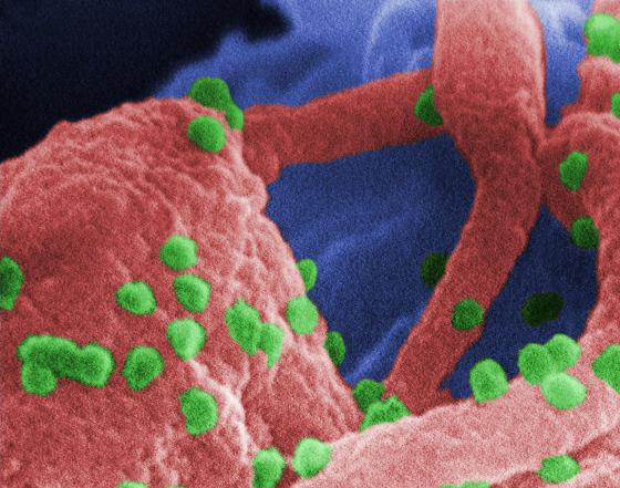
A retrovirus, the Human Immunodeficiency Virus (HIV) was identified in 1983 as the pathogen responsible for the Acquired Immunodeficiency Syndrome (AIDS). AIDS is characterized by changes in the population of T-cell lymphocytes that play a key role in the immune defense system. In the infected individual, the virus causes a depletion of T-cells, called “T-helper cells”, which leaves these patients susceptible to opportunistic infections, and certain malignancies. Credit: CDC/ C. Goldsmith, P. Feorino, E. L. Palmer, W. R. McManus

How many people does HIV/AIDS affect?

AIDS is the sixth leading cause of death among people aged 25 - 44 in the United States. This is an improvement since it was the number one killer in 1995.
At the end of 2010, an estimated 91,500 people in the UK were living with HIV. Of these, around 1 in 4 (22,000 in total) did not know they were infected.
The World Health Organization (WHO) estimates that around 34 million people in the world are living with HIV. The virus is particularly widespread in sub-Saharan African countries, such as South Africa, Zimbabwe and Mozambique.

Cause of AIDS

AIDS is caused by HIV infection. The virus attacks the immune system leaving the individual susceptible to life-threatening infections and cancers. Common bacteria, yeast, parasites, and viruses that usually do not cause serious disease in people with healthy immune systems can turn deadly for AIDS patients.

How is HIV transmitted

HIV is found in all the body fluids including saliva, nervous system tissue and spinal fluid, blood, semen, pre-seminal fluid, which is the liquid that comes out before ejaculation, vaginal secretions, tears and breast milk. Only blood, semen, and breast milk have been shown to transmit infection to others.
The virus is transmitted by sexual contact including unprotected oral, vaginal, and anal sex and via transfusion of contaminated blood that contains HIV.
Another mode of transmission is sharing needles or injections with HIV infected individuals.
A pregnant woman can transmit the virus to her unborn baby through their shared blood circulation, or a nursing mother can transmit it to her baby in her breast milk.
HIV infection does not spread by casual contact, mosquitoes, touching or hugging.

Who is at risk?

Those at highest risk include injection drug users who share needles, babies born to mothers with HIV (especially if the mother had not received anti- HIV therapy during pregnancy), those engaging in unprotected vaginal or anal sex with HIV positive individuals, and those who received blood transfusions or clotting products between 1977 and 1985 (before screening for HIV became standard practice).

Symptoms of HIV/AIDS

HIV infection may cause no symptoms for a decade or longer. At this stage carriers may transmit the infection to others unknowingly. If the infection is not detected and treated, the immune system gradually weakens and AIDS develops.
Acute HIV infection takes a few weeks to months to become a non-symptomatic HIV infection. Then it becomes early symptomatic HIV infection and later it progresses to AIDS.

How is the progress of the disease marked?

With advancing HIV infection the blood shows higher viral load and CD4 T-cell count drops below 200 cells/mm3. CD4 cells are a type of T cell. T cells are cells of the immune system. They are also called "helper cells."
There is a small group of patients who develop AIDS very slowly, or never at all. These patients are called nonprogressors, and many seem to have a genetic difference that prevents the virus from significantly damaging their immune system.

Opportunistic infections

These are infections that normally do not affect an individual with a healthy immune system but AIDS patients are susceptible to these infections. These include viral infections like:
  • herpes simplex virus
  • herpes zoster infection
  • cancers like Kaposi sarcoma, non-Hodgkin’s lymphoma
  • fungal infections like candidiasis
  • bacterial infections like tuberculosis
Other infections include Bacillary angiomatosis, Candida esophagitis, Pneumocystic jiroveci pneumonia, AIDS dementia, Cryptosporidium diarrhea, cryptococcal meningigits and Toxoplasma encephalitis.

Treatment of AIDS

There is no cure for AIDS once it develops. There are agents available that can help keep symptoms at bay and improve the quality and length of life for those who have already developed symptoms.
Drugs against HIV include antiretroviral therapy. These prevent the replication of the HIV virus in the body. A combination of several antiretroviral drugs, called highly active antiretroviral therapy (HAART), has been very effective in reducing the number of HIV particles in the bloodstream. Preventing the virus from replicating can improve T-cell counts or CD4 cell counts and help the immune system recover from the HIV infection.
Medicines are also prescribed to prevent opportunistic infections if the CD4 counts are low.

Outcome of HIV

AIDS is almost always fatal without treatment. HAART however has dramatically increased the amount of time people with HIV remain alive.

Prevention of HIV

Safe sex measures with use of condoms, shunning use of illicit drugs or shared needles or syringes, avoidance of contact with blood and fluids by wearing protective clothing, masks, and goggles etc. helps prevent transmission.
HIV-positive women who wish to become pregnant may need therapy while they are pregnant to prevent transmission to their babies. The Public Health Service recommends that HIV-infected women in the United States avoid breastfeeding to prevent transmitting HIV to their infants through breast milk.

What is a Blood Transfusion?


Blood transfusion is the process of transferring blood or blood-based products from one person into the circulatory system of another. Blood transfusions can be life-saving in some situations, such as massive blood loss due to trauma, or can be used to replace blood lost during surgery.
Blood transfusions may also be used to treat a severe anaemia or thrombocytopenia caused by a blood disease. People suffering from hemophilia or sickle-cell disease may require frequent blood transfusions. Early transfusions used whole blood, but modern medical practice commonly uses only components of the blood.
Blood transfusions can be grouped into two main types depending on their source:
  • ''Homologous transfusions'', or transfusions using the stored blood of others. These are often called ''Allogeneic'' instead of homologous.
  • ''Autologous transfusions'', or transfusions using the patient's own stored blood.
Donor units of blood must be kept refrigerated to prevent bacterial growth and to slow cellular metabolism. The transfusion must begin within 30 minutes after the unit has been taken out of controlled storage.
Blood can only be administered intravenously. It therefore requires the insertion of a cannula of suitable caliber.
Before the blood is administered, the personal details of the patient are matched with the blood to be transfused, to minimize risk of transfusion reactions. Clerical error is a significant source of transfusion reactions and attempts have been made to build redundancy into the matching process that takes place at the bedside.
A unit (up to 500 ml) is typically administered over 4 hours. In patients at risk of congestive heart failure, many doctors administer a diuretic to prevent fluid overload, a condition called Transfusion Associated Circulatory Overload or TACO. Acetaminophen and/or an antihistamine such as diphenhydramine are sometimes given before the transfusion to prevent other types of transfusion reactions.
Blood is most commonly donated as whole blood by inserting a catheter into a vein and collecting it in a plastic bag (mixed with anticoagulant) via gravity. Collected blood is then separated into components to make the best use of it. Aside from red blood cells, plasma, and platelets, the resulting blood component products also include albumin protein, clotting factor concentrates, cryoprecipitate, fibrinogen concentrate, and immunoglobulins (antibodies). Red cells, plasma and platelets can also be donated individually via a more complex process called apheresis.
In developed countries, donations are usually anonymous to the recipient, but products in a blood bank are always individually traceable through the whole cycle of donation, testing, separation into components, storage, and administration to the recipient. This enables management and investigation of any suspected transfusion related disease transmission or transfusion reaction. In developing countries the donor is sometimes specifically recruited by or for the recipient, typically a family member, and the donation immediately before the transfusion.

Risks to the recipient

There are risks associated with receiving a blood transfusion, and these must be balanced against the benefit which is expected. The most common adverse reaction to a blood transfusion is a ''febrile non-hemolytic transfusion reaction'', which consists of a fever which resolves on its own and causes no lasting problems or side effects.
Hemolytic reactions include chills, headache, backache, dyspnea, cyanosis, chest pain, tachycardia and hypotension.
Blood products can rarely be contaminated with bacteria; the risk of severe bacterial infection and sepsis is estimated, as of 2002, at about 1 in 50,000 platelet transfusions, and 1 in 500,000 red blood cell transfusions.
There is a risk that a given blood transfusion will transmit a viral infection to its recipient. As of 2006, the risk of acquiring hepatitis B via blood transfusion in the United States is about 1 in 250,000 units transfused, and the risk of acquiring HIV or hepatitis C in the U.S. via a blood transfusion is estimated at 1 per 2 million units transfused. These risks were much higher in the past before the advent of second and third generation tests for transfusion transmitted diseases. The implementation of Nucleic Acid Testing or "NAT" in the early 00's has further reduced risks, and confirmed viral infections by blood transfusion are extremely rare in the developed world.
Transfusion-associated acute lung injury (TRALI) is an increasingly recognized adverse event associated with blood transfusion. TRALI is a syndrome of acute respiratory distress, often associated with fever, non-cardiogenic pulmonary edema, and hypotension, which may occur as often as 1 in 2000 transfusions. Symptoms can range from mild to life-threatening, but most patients recover fully within 96 hours, and the mortality rate from this condition is less than 10%.. Although the cause of TRALI is not clear, it has been consistently associated with anti HLA antibodies. Because anti HLA strongly correlate with pregnancy, several transfusion organisations (Blood and Tissues Bank of Cantabria, Spain, National Health Service in Britain) have decided to use only plasma from men for transfusion.
Other risks associated with receiving a blood transfusion include volume overload, iron overload (with multiple red blood cell transfusions), transfusion-associated graft-vs.-host disease, anaphylactic reactions (in people with IgA deficiency), and acute hemolytic reactions (most commonly due to the administration of mismatched blood types).

Transformation from one type to another

Scientists working at the University of Copenhagen reported in the journal Nature Biotechnology in April 2007 of discovering enzymes, which potentially enable blood from groups A, B and AB to be converted into group O. These enzymes do not affect the Rh group of the blood.

Objections to blood transfusion

Objections to blood transfusions may arise for personal, medical, or religious reasons. For example, Jehovah's Witnesses object to blood transfusion primarily on religious grounds - they believe that blood is sacred; although they have also highlighted possible complications associated with transfusion.

Storing Breast Milk


It is important to know the guidelines for storing breast milk properly so that you always give your baby fresh milk. Any container used to store milk should be clean and sterile. Always try to leave an inch or so from the milk to the top of the container since frozen milk expands. After pumping your milk, it is helpful to label the storage container. Always use the oldest dated milk first. Colostrum, or the first milk expressed in the first few days after delivery, can be stored at room temperature for up to 12 hours. Mature milk, or breast milk that comes in six days after the birth of your baby can be stored in the following ways:

At Room Temperature:

  • At 60 degrees Fahrenheit for 24 hours (15.5 Celcius)
  • At 66-72 degrees Fahrenheit for 10 hours (19-22 Celcius)
  • At 79 degrees Fahrenheit for 4-6 hours (26 Celcius)
  • At 86-100 degrees Fahrenheit for 4 hours (30-37 Celcius)

In the Refrigerator:

  • At 32-39 degrees Fahrenheit for up to 8 days (0-4 Celcius)

In the Freezer*:

  • In a freezer compartment contained within the refrigerator for up to 2 weeks
  • In a self-contained freezer, either on top of or on the side of the refrigerator for 3 - 4 months
  • In a deep freezer with a constant 0 degrees Fahrenheit for 6 months or longer (-17 Celcius)
*It is helpful to freeze the milk in small amounts, such as 2 to 4 ounce servings, so there is less waste and you can choose the amount of milk depending on the baby's hunger.

Bottles and Containers

You can store breast milk in bottles that fit directly onto your breast pump. After pumping, simply remove the pumping tubing, cover with the bottle lid, label the milk, and put it in the refrigerator. Many breast pump carrying cases also come with built-in, cooler-type compartments for storing ice pack and/or the freshly pumped bottles of milk. If used correctly, these do stay cold enough to leave your pumped milk in until you can get home to store the milk in the refrigerator or freezer.
Research is conflicting about the advantages and disadvantages of storing milk in glass versus plastic. However, glass bottles or containers are best for freezing breast milk because it offers the most protection from contamination. The second choice is clear, hard plastic, and the last choice is the cloudy hard plastic containers. Wait to tighten the caps or lids until the milk is completely frozen.

Storage Bags

If you want to freeze your breast milk in bags, you can purchase storage bags that fit directly onto your breast pump and that are made for freezing milk. They are pre-sterilized, thick, have an area for labeling, and seal easily. After pumping, simply remove the pumping tubing, fold the bag over, making sure all air is out of the bag, and seal it. Make sure to label the bag with the date before freezing. When you want to use the milk, you can cut the storage bag with sterile scissors. If the storage bag has a built-in pouring spout, it is easy to pour the milk into a bottle. Other storage bags can be used in the kind of bottle that uses disposable liners, so there is no need to transfer the milk.

Thawing and Handling Stored Breast Milk

It is normal for stored breast milk to separate in its container into two parts, what looks like cream and then a lighter colored milk. Some human milk also varies in color and can be blueish, yellowish, or brownish. Just gently shake the milk before feeding to mix it back together.
Breast milk doesn't take long to thaw or warm up. Never place a bottle or bag of breast milk in the microwave. Milk doesn't heat uniformly in the microwave, so you won't have control over the temperature and could burn your baby. All you have to do is hold the bottle or frozen bag of milk under cool and then warm water for a few minutes. If warm running water is not available, you can heat up a pan of water on the stove. Remove the pan from the heat and place the container into the warm water. Never warm the container directly on the stove. Shake the milk, then test it on your wrist to see if it's warm enough for your baby.

Semen - What is Semen?


Semen is a greyish white bodily fluid that is secreted by the gonads of male animals. It carries sperm or the spermatozoa and fructose and other enzymes that help the sperm to survive to facilitate successful fertilization.
The whitish opalescence is due to the large amount of protein that it contains and its slightly turbid appearance is due to the spermatozoa contained within it.

Process of ejaculation

Semen is released during the process of ejaculation and is processed in the seminal vesicle in the pelvis, which is where it is produced.

How does ejaculation occur?

Ejaculation is controlled by the central nervous system and occurs when there is friction on the genitalia and other forms of sexual stimulation. The stimuli lead to impulses that are sent up the spinal cord and into the brain.

Two phases of ejaculation

Ejaculation has two phases:
Phase 1: emission in which the vas deferens (the tubes that store and transport sperm from the testes) contract to squeeze the sperm toward the base of the penis through the prostate gland and into the urethra. The seminal vesicles release their part of the semen that combine with the sperm. The ejaculation is unstoppable at this stage.
Phase 2: ejaculation in which the muscles at the base of the penis and urethra contract. This leads to forcing the semen out of the penis (ejaculation and orgasm) and this phase also has a bladder neck contraction. The bladder neck contracts to prevent the back flow of the semen into the urinary tract. Dry orgasm can occur even without delivery of semen (ejaculation) from the penis. Erection declines normally following ejaculation.

Semen composition

The semen travels through the ejaculatory ducts and mixes with fluids from the seminal vesicles, the prostrate, and the bulbourethral glands.
The seminal vesicles produce a viscous, fructose-rich fluid forming around 65-70% of the semen base.
The white color of the semen is due to secretion from the prostate glands containing enzymes, citric acid, lipids, and acid phosphatase. This forms around 25-30% of the semen base.
At each ejaculation around 200-500 million sperms are released by the testes. This forms about 2-5% of the semen composition.
Apart from these, the bulbourethral glands produce a clear secretion. This helps in mobility of the sperm cells in the vagina and cervix. The glands’ secretion contribute less than 1% to the overall semen composition.
The semen comprises of:
  • fructose
  • ascorbic acid
  • zinc
  • cholesterol
  • protein
  • calcium
  • chlorine
  • blood group antigens
  • citric acid
  • DNA
  • Magnesium
  • vitamin B12
  • phosphorus
  • sodium
  • potassium
  • uric acid
  • lactic acid
  • nitrogen
  • other nutrients

Semen per ejaculation

Ejaculation is a complex process and the compositions of the final semen come together in the posterior urethra and only become mixed after ejaculation is complete.
The volume of semen released per ejaculate varies. Approximately an average around 3.4 milliliters is ejaculated at one time. It can be as high as 4.99 milliliters or as low as 2.3 milliliters.
If there is a prolonged gap between ejaculations, the number of sperm in the semen increases but there is no overall increase in the semen.

What is the Nervous System?


The nervous system is a complex network of nerves and cells that carry messages to and from the brain and spinal cord to various parts of the body.
The nervous system includes both the Central nervous system and Peripheral nervous system. The Central nervous system is made up of the brain and spinal cord and The Peripheral nervous system is made up of the Somatic and the Autonomic nervous systems.

The Central nervous system

The central nervous system is divided into two major parts: the brain and the spinal cord.

The brain

The brain lies within the skull and is shaped like a mushroom.  The brain consists of four principal parts:
  • the brain stem
  • the cerebrum
  • the cerebellum
  • the diencephalon
The brain weighs approximately 1.3 to 1.4 kg. It has nerve cells called the neurons and supporting cells called the glia.
There are two types of matter in the brain:  grey matter and white matter.  Grey matter receives and stores impulses.  Cell bodies of neurons and neuroglia are in the grey matter.  White matter in the brain carries impulses to and from grey matter.  It consists of the nerve fibers (axons).

The brain stem

The brain stem is also known as the Medulla oblongata. It is located between the pons and the spinal cord and is only about one inch long.

The cerebrum

The cerebrum forms the bulk of the brain and is supported on the brain stem.  The cerebrum is divided into two hemispheres.  Each hemisphere controls the activities of the side of the body opposite that hemisphere. 
The hemispheres are further divided into four lobes:
  • Frontal lobe
  • Temporal lobes
  • Parietal lobe
  • Occipital lobe

The cerebellum

This is located behind and below the cerebrum. 

The diencephalon

The diencephalon is also known as the fore brain stem. It includes the thalamus andhypothalamus. The thalamus is where sensory and other impulses go and coalesce.
The hypothalamus is a smaller part of the diencephalon

Other parts of the brain

Other parts of the brain include the midbrain and the pons:
  • the midbrain provides conduction pathways to and from higher and lower centers
  • the pons acts as a pathway to higher structures;  it contains conduction pathways between the medulla and higher brain centers

The spinal cord

The spinal cord is along tube like structure which extends from the brain. The spinal cord is composed of a series of 31 segments.  A pair of spinal nerves comes out of each segment.  The region of the spinal cord from which a pair of spinal nerves originates is called the spinal segment.  Both motor and sensory nerves are located in the spinal cord.
The spinal cord is about 43 cm long in adult women and 45 cm long in adult men and weighs about 35-40 grams. It lies within the vertebral column, the collection of bones (back bone).

Other parts of the central nervous system

The meninges are three layers or membranes that cover the brain and the spinal cord.  The outermost layer is the dura mater.  The middle layer is the arachnoid, and the innermost layer is the pia mater. The meninges offer protection to the brain and the spinal cord by acting as a barrier against bacteria and other microorganisms.
The Cerebrospinal Fluid (CSF) circulates around the brain and spinal cord. It protects and nourishes the brain and spinal cord.

Neurons

The neuron is the basic unit in the nervous system. It is a specialized conductor cell that receives and transmits electrochemical nerve impulses. A typical neuron has a cell body and long arms that conduct impulses from one body part to another body part.
There are three different parts of the neuron:
  • the cell body
  • dendrites
  • axon

Cell body of a neuron

The cell body is like any other cell with a nucleus or control center. 

Dendrites

The cell body has several highly branched, thick extensions that appear like cables and are called dendrites.  The exception is a sensory neuron that has a single, long dendrite instead of many dendrites.  Motor neurons have multiple thick dendrites. The dendrite's function is to carry a nerve impulse into the cell body.

Axon

An axon is a long, thin process that carries impulses away from the cell body to another neuron or tissue.  There is usually only one axon per neuron. 

Myelin Sheath

The neuron is covered with the Myelin Sheath or Schwann Cells. These are white segmented covering around axons and dendrites of many peripheral neurons. The covering is continuous along the axons or dendrites except at the point of termination and at the nodes of Ranvier.
The neurilemma is the layer of Schwann cells with a nucleus. Its function is to allow damaged nerves to regenerate.  Nerves in the brain and spinal cord do not have a neurilemma and, therefore cannot recover when damaged.

Types of neuron

Neurons in the body can be classified according to structure and function. According to structure neurons may be multipolar neurons, bipolar neurons, and unipolar neurons:
  • Multipolar neurons have one axon and several dendrites. These are common in the brain and spinal cord
  • Bipolar neurons have one axon and one dendrite.  These are seen in the retina of the eye, the inner ear, and the olfactory (smell) area. 
  • Unipolar neurons have one process extending from the cell body. The one process divides with one part acting as an axon and the other part functioning as dendrite. These are seen in the spinal cord.

The Peripheral nervous system

The Peripheral nervous system is made up of two parts:
  • Somatic nervous system
  • Autonomic nervous system

Somatic nervous system

The somatic nervous system consists of peripheral nerve fibers that pick up sensory information or sensations from the peripheral or distant organs (those away from the brain like limbs) and carry them to the central nervous system.
These also consist of motor nerve fibers that come out of the brain and take the messages for movement and necessary action to the skeletal muscles. For example, on touching a hot object the sensory nerves carry information about the heat to the brain, which in turn, via the motor nerves, tells the muscles of the hand to withdraw it immediately.
The whole process takes less than a second to happen. The cell body of the neuron that carries the information often lies within the brain or spinal cord and projects directly to a skeletal muscle.

Autonomic Nervous System

Another part of the nervous system is the Autonomic Nervous System. It has three parts:
  • the sympathetic nervous system
  • the parasympathetic nervous system
  • the enteric nervous system
This nervous system controls the nerves of the inner organs of the body on which humans have no conscious control. This includes the heartbeat, digestion, breathing (except conscious breathing) etc.
The nerves of the autonomic nervous system enervate the smooth involuntary muscles of the (internal organs) and glands and cause them to function and secrete their enzymes etc.
The Enteric nervous system is the third part of the autonomic nervous system. The enteric nervous system is a complex network of nerve fibers that innervate the organs within the abdomen like the gastrointestinal tract, pancreas, gall bladder etc. It contains nearly 100 million nerves.

Neurons in the peripheral nervous system

The smallest worker in the nervous system is the neuron. For each of the chain of impulses there is one preganglionic neuron, or one before the cell body or ganglion, that is like a central controlling body for numerous neurons going out peripherally.
The preganglionic neuron is located in either the brain or the spinal cord. In the autonomic nervous system this preganglionic neuron projects to an autonomic ganglion. The postganglionic neuron then projects to the target organ.
In the somatic nervous system there is only one neuron between the central nervous system and the target organ while the autonomic nervous system uses two neurons.

Basic Virus


What is a Virus?

Viruses are tiny organisms that may lead to mild to severe illnesses in humans, animals and plants. This may include flu or a cold to something more life threatening likeHIV/AIDS.

How big are viruses?

The virus particles are 100 times smaller than a single bacteria cell. The bacterial cell alone is more than 10 times smaller than a human cell and a human cell is 10 times smaller than the diameter of a single human hair.

Are viruses alive?

Viruses by themselves are not alive. They cannot grow or multiply on their own and need to enter a human or animal cell and take over the cell to help them multiply. These viruses may also infect bacterial cells.
The virus particle or the virions attack the cell and take over its machinery to carry out their own life processes of multiplication and growth. An infected cell will produce viral particles instead of its usual products.

Structure of a virus

A virion (virus particle) has three main parts:
  • Nucleic acid – this is the core of the virus with the DNA or RNA (deoxyribonucleic acid and ribonucleic acid respectively). The DNA or RNA holds all of the information for the virus and that makes it unique and helps it multiply.
  • Protein Coat (capsid) – This is covering over the nucleic acid that protects it.
Lipid membrane (envelope) – this covers the capsid. Many viruses do not have this envelope and are called naked viruses.

Receptors

Viruses are not simply taken into cells. They must first attach to a receptor on the cell surface. Each virus has its specific receptor, usually a vital component of the cell surface. It is the distribution of these receptor molecules on host cells that determines the cell-preference of viruses. For example, the cold and flu virus prefers the mucus lining cells of the lungs and the airways.

How do viruses infect?

Viruses do not have the chemical machinery needed to survive on their own. They, thus seek out host cells in which they can multiply. These viruses enter the body from the environment or other individuals from soil to water to air via nose, mouth, or any breaks in the skin and seek a cell to infect.
A cold or flu virus for example will target cells that line the respiratory (i.e. the lungs) or digestive (i.e. the stomach) tracts. The HIV (human immunodeficiency virus) that causes AIDS attacks the T-cells (a type of white blood cell that fights infection and disease) of the immune system.

Life cycle of a basic virus

There are a few basic steps that all infecting viruses follow and these are called the lytic cycle. These include:
  1. A virus particle attaches to a host cell. This is called the process of adsorption
  2. The particle injects its DNA or RNA into the host cell called entry.
  3. The invading DNA or RNA takes over the cell and recruits the host’s enzymes
  4. The cellular enzymes start making new virus particles called replication
  5. The particles of the virus created by the cell come together to form new viruses. This is called assembly
  6. The newly formed viruses kill the cell so that they may break free and search for a new host cell. This is called release.

What is Microscopy?


Microscopy is the technical field of usnig microscopes to view samples and objects that cannot be seen with the unaided eye (objects that are not within the resolution range of the normal eye). There are three well-known branches of microscopy: optical, electron, and scanning probe microscopy.
Optical and electron microscopy involve the diffraction, reflection, or refraction of electromagnetic radiation/electron beams interacting with thespecimen, and the subsequent collection of this scattered radiation or another signal in order to create an image. This process may be carried out by wide-field irradiation of the sample (for example standard light microscopy and transmission electron microscopy) or by scanning of a fine beam over the sample (for example confocal laser scanning microscopy and scanning electron microscopy). Scanning probe microscopy involves the interaction of a scanning probe with the surface of the object of interest. The development of microscopy revolutionized biology and remains an essential technique in the life and physical sciences.

সোমবার, ১৫ অক্টোবর, ২০১২

Pneumonia


What is pneumonia?

Pneumonia is a lung infection that can make you very sick. You may cough, run a fever, and have a hard time breathing. For most people,pneumonia can be treated at home. It often clears up in 2 to 3 weeks. But older adults, babies, and people with other diseases can become very ill. They may need to be in the hospital.
You can get pneumonia in your daily life, such as at school or work. This is called community-associated pneumonia. You can also get it when you are in a hospital or nursing home. This is called healthcare-associated pneumonia. It may be more severe because you already are ill. This topic focuses on pneumonia you get in your daily life.

What causes pneumonia?

Germs called bacteria or viruses usually cause pneumonia.
Pneumonia usually starts when you breathe the germs into your lungs. You may be more likely to get the disease after having a cold or the flu. These illnesses make it hard for your lungs to fight infection, so it is easier to get pneumonia. Having a long-term, or chronic, disease like asthma, heart disease, cancer, or diabetes also makes you more likely to get pneumonia.

What are the symptoms?

Symptoms of pneumonia caused by bacteria usually come on quickly. They may include:
  • Cough. You will likely cough up mucus (sputum) from your lungs. Mucus may be rusty or green or tinged with blood.
  • Fever.
  • Fast breathing and feeling short of breath.
  • Shaking and "teeth-chattering" chills. You may have this only one time or many times.
  • Chest pain that often feels worse when you cough or breathe in.
  • Fast heartbeat.
  • Feeling very tired or feeling very weak.
  • Nausea and vomiting.
  • Diarrhea.
When you have mild symptoms, your doctor may call this "walking pneumonia."
Older adults may have different, fewer, or milder symptoms. They may not have a fever. Or they may have a cough but not bring up mucus. The main sign of pneumonia in older adults may be a change in how well they think. Confusion ordelirium is common. Or, if they already have a lung disease, that disease may get worse.
Symptoms caused by viruses are the same as those caused by bacteria. But they may come on slowly and often are not as obvious or as bad.

How is pneumonia diagnosed?

Your doctor will ask you about your symptoms and do a physical exam. He or she may order a chest X-ray and a blood test. This is usually enough for your doctor to know if you have pneumonia. You may need more tests if you have bad symptoms, are an older adult, or have other health problems. In general, the sicker you are, the more tests you will have.

Diabetes


Diabetes is a lifelong condition in which sugar (glucose) remains in the blood rather than entering the body’s cells to be used for energy. This results in persistently high blood sugar, which, over time, can damage many body systems.
Symptoms of diabetes include increased thirst and frequent urination (especially at night); unexplained increase in appetite; unexplained weight loss; fatigue; erection problems; blurred vision; and tingling, burning, or numbness in the hands or feet.
People who have diabetes are at increased risk for many serious health problems, including hardening of the arteries (atherosclerosis) and heart problems, eye problems that can lead to blindness, circulation and nerve problems, and kidney disease and kidney failure.
Pregnant women with uncontrolled diabetes have an increased risk of miscarriage and birth defects.
Diabetes is treated with diet and lifestyle changes and with medicines (such as insulin or oral medicines). If blood sugar levels are kept within the recommended range, the risk for many complications from diabetes decreases.

Asthma


Asthma is a long-term (chronic) disease of the respiratory system that causes inflammation and spasm or tightening in the bronchial tubes, which carry air to the lungs. The inflammation causes periodic episodes of difficulty breathing, wheezing, chest tightness, and coughing.
Although asthma cannot be cured, most people can control their asthma by following a plan, avoiding triggers, and taking medicine.
Asthma often begins during childhood and may last throughout a person's life.
The cause of asthma is not clearly known. It is more common in people who also have allergies.

Influenza (flu)


Influenza (flu) is a viral upper respiratory illness that comes on suddenly, causing a person to feel very sick with symptoms such as fever, body aches, headache, fatigue, loss of appetite, and a dry cough or sore, dry throat. Home treatment to reduce discomfort is usually all that is needed.
In some cases, the flu can lead to complications, such as bacterial pneumonia. People who are at high risk for complications include young children up to 5 years of age, adults age 65 and older, and all those who have certain health conditions, such as chronic heart or lung problems or immune system disorders.
Yearly immunization with the inactivated flu vaccine (flu shot) or the nasal spray vaccine (FluMist) helps reduce the risk of getting the flu and makes symptoms less severe for people who do get the flu.

Lungs

Picture of the Human Lungs

Picture of the Lungs

The lungs are a pair of spongy, air-filled organs located on either side of the chest (thorax). The trachea (windpipe) conducts inhaled air into the lungs through its tubular branches, called bronchi. The bronchi then divide into smaller and smaller branches (bronchioles), finally becoming microscopic.
The bronchioles eventually end in clusters of microscopic air sacs called alveoli. In the alveoli, oxygen from the air is absorbed into the blood. Carbon dioxide, a waste product of metabolism, travels from the blood to the alveoli, where it can be exhaled. Between the alveoli is a thin layer of cells called the interstitium, which contains blood vessels and cells that help support the alveoli.
The lungs are covered by a thin tissue layer called the pleura. The same kind of thin tissue lines the inside of the chest cavity -- also called pleura. A thin layer of fluid acts as a lubricant allowing the lungs to slip smoothly as they expand and contract with each breath.

Pneumonia


Pneumonia is an inflammation of the lungs most often caused by infection with bacteria or a virus. Pneumonia can make it hard to breathe because the lungs have to work harder to get enough oxygen into the bloodstream.
Symptoms of pneumonia caused by bacteria often begin suddenly and may follow an upper respiratory infection, such as influenza (flu) or a cold. Common symptoms include fever, a cough that often produces colored mucus (sputum) from the lungs, and rapid, often shallow breathing.
Older adults may have different, fewer, or milder symptoms. The major sign of pneumonia in older adults may be a change in how well they think (confusion or delirium) or a worsening of a lung disease they already have.
Symptoms of pneumonia not caused by bacteria (nonbacterial) include fever, cough, and shortness of breath, and there may be little mucus production.
Antibiotics are used to treat pneumonia caused by bacteria.

রবিবার, ১৪ অক্টোবর, ২০১২

Vertebrates


Vertebrates

Vertebrates - Back boned animals:
Not only vertebrates are back boned animals because vertebrata is not the phylum of back boned animals. Chordata is the phylum of back boned animals and vertebrata is the sub-phylum of chordata. Here, back bone is generally considered as notochord. This notochord is strong and rigid to give good support to chordate animals. But in vertebrates, it was modified as jointed backbone which is called as 'vertebrae'. Animals those possess vertebrae is called as vertebrates.
Modern and major classification of vertebrates:
As per the modern day of classification, there are only five classes of vertebrates. They are
  • Pisces (Fishes)
  • Amphibia (Amphibians such as Frogs and toads,etc.)
  • Reptilia (Crocodiles, snakes, Dinosaurs, turtles, etc.)
  • Aves (All birds including penguins & excluding bats, etc.)
  • Mammalia (Rats, Rabbits, Elephants, Whales, Bats, Human beings, etc.)
Pisces - Aquatic animals:
Pisces is the class where all aquatic animals such as fishes excluding dolphins, whales and some aquatic amphibians and reptiles. Fishes originate in the Devonian period which is considered as the "Age of fishes". Fishes again classified into:
  • Agnatha (Jawless fishes)
  • Chondrichthyes (Cartilaginous fishes such as shark, ray, etc.)
  • Osteichthyes (Bony fishes such as Catfish, Tilapia, Carps, etc.)
We can see that very common adaptation acquired by fishes for aquatic life. They are
  • Streamlined body,
  • Gills for respiring dissolved oxygen in water,
  • Fins present at suitable places for effective swimming.
Amphibians - Animals evolved from water to land:
It is believed that the amphibians evolved from labyrinthodonts which are the transitional animal group between fish and amphibians. The most familiar animals found in amphibians are frogs and toads, etc.
The adaptation acquired by amphibians is:
  • Cutaneous respiration when the animals found inside the water,
  • Nictitating membrane found in the eye to get clear view inside the water.
  • Webbed foot to give effective swimming.
Reptiles - Amazing animals:
Yes, reptiles are very amazing because the size range of animals also very amazing, as it ranges from very small lizard to huge dinosaurs. You can see variety of adaptations in this group because it habituated in water, in land, in damps, in deserts, etc.
Birds - Aerial animals:
Even in invertebrates, insects exhibit flight, but the birds shows exact flight adaptation, that's why it is very perfect aerial animals. The flight adaptations are:
  • Reduced bone marrow to reduce bone weight.
  • Pored bones
  • Air sacs,
  • Bursa of fabricius,
  • Feathers & wings, etc.
Mammals - Milk feeding animals:
The striking characteristic of mammals is feeding milk to their young ones. Based on their habits and adaptations, it is diverged into various groups such as
  • Burrowing rats and moles,
  • Flying bats,
  • Fast running cheetahs,
  • Long leaping kangaroo,
  • Aquatic whales and dolphins, etc.

Classify Pisces upto sub-class giving salient features and examples


The super class Pisces (L., Piscis, fish) includes all the fishes which are essentially aquatic forms with paired fins for swimming and gills for locomotion. About 40,000 species of fishes are known. Various workers have provided different schemes of their classification.
However, no classification had been universally accepted because of the confusion due to staggering numbers of fishes and great diversity in their shape, size, habits and habitat.
According to Parker and Haswell (1960) the super class Pisces if sub-divided into three classes.
CLASS-I-PLACODERMI
(i) These include all extinct fishes found from early Devonian to Permian period.
(ii) Body heavily armoured with bony scales or plates.
(iii) Primitive jaws with teeth.
(iv) Paired or unpaired fins were present.
(v) Skeleton bony.
(vi) Notochord persistent throughout life.
(vii) They are immediate ancestors of astracoderms.
The class placodermi includes six subclasses.
(i) Sub-class – I – Acanthodii, Ex: Climatius
(ii) Sub-class – II – Arthrodin, Ex: Cocoosteus
(iii) Sub-class – III – Petalichthyda, Ex: Macropetalch
(iv) Sub-class – IV – Antiarchi, Ex: Pterich thyodes
(v) Sub-class – V – Rhenanida, Ex: Gemuendina
(vi) Sub-class – VI – Palaeospondylia, Ex: Palaeospondylus
CLASS-II – CHONDRICHTHYES:
(i) Mostly marine and predaceous
(ii) Body fusiform or spindle shaped.
(iii) Endeskeleton is cartilaginous
(iv) Skin with placoid scales.
(v) Fins both median and paired all, supported by fin rays. Pelvic fin bears claspers in males.
(vi) Notochord tail fin heterocereal persistant. Vertebrae complete and separate from notochord.
(vii) Mouth ventral in position, jaws p[resent. Teeth are modified placoid scales.
(viii) Digestive system complete, stomach J-shaped, intestine with spiral valves.
(ix) Gill slits separate, 5-7 pairs, laterally placed, without operculum.
(x) Air bladder and lungs absent.
(xi) Heart 2-chambered, poikilothermous.
(xii) Kidneys opisthonephrii, Ureotelic.
(xiii) Sexes separate, Gonads paired, fertilization internal, oviparous or ovoviviparous. It includes two sub-classes.
1. Sub-class-I-SELACHII (Elasmorbranchii
(i) Multiple gill slits on either side protected but individual skin flaps.
(ii) A spiracle behind each eye.
(iii) Cloaca present.
Ex: Scolodon (shark), Torpedo (Electric ray) pristis (saw fish)
(ii) sub-class-II – HOLOCEPHALUI
(i) Single gill opening on either side covered by a fleshy operculum.
(ii) No spiracles, cloaca and sales.
(iii) Single nasal opening.
Ex: Chimaeras or Rat fishes.
Class-III- OSTEICHTHYES (Teleostomi)
(i) Body is spindle shaped.
(ii) Median and paired fins are present.
(iii) Tail fin is usually Homocercal.
(iv) Endoskeleton is partly or wholly bony.
(v) Skin covered by 3 types of dermal scales ganoid, cycloid or ctenoid. Some without scales. No placoid scales.
(vi) Mouth terminal or subterminal Jaws usually with teeth. Cloaca lacking, anus present.
(vii) Gills are covered by a common operculum on either side.
(viii) An air (swim) bladder often present with or without duct connected t pharynx.
(ix) Adult kidneys mesonephric.
(x) Well developed lateral line system. Internal ear with 3 semicircular canals.
(xi) Sexes separate, Gonads paired fertilization usually external.
(xii) Mostly oviparous, rarely ovoviviparous or viviparous.
This subclass includes two sub-classes.
Sub-class-I – Sarcopterygii
(i) Paired fins are leg-like or lobed with a fleshy, bony central axis covered by scales.
(ii) Internal nares present.
(iii) All are fresh water forms.
Ex: - Latimeria (living fossil)
Protopterus & Lepidosiren
(Lung-fishes)
Sub-class II – Actinoptergii
(i) Paired fins thin, broad without fleshy basal lobes.
(ii) Double external nares are present
(iii) Popularly called ray-finned fish.
This subclass is divided into the infraclasses or super orders
(i) Chondrostei Ex: - Polypterus
(ii) Holostei Ex: - Lepidosteus
(iii) Telestoei Ex: - Labeo, Catla