বৃহস্পতিবার, ৩১ মে, ২০১২

Define carbohydrate. Write its classification.


Carbohydrates: Carbohydrates are organic compounds that contain C,H,O atoms. Carbohydrates are mainly consist of  monosaccharids or the compound of mono sugar of various lengths. The word “saccharide” is derived from the geek word “sakkharon”, meaning sugar.

Carbohydrates may be defined chemically as aldehyde or ketone derivatives of polyhydric (more than one –OH group) alcohols or as compounds that yield these derivatives on hydrolysis.
                                               –Harper

The general formula of the carbohydrates is Cn(H2O)n. As the ratio of H and O in carbohydrate is 2:1 same as water, so they are often considered as “hydrated carbon”. Some carbohydrates also contain nitrogen, phosphorus or sulfur.


Classification of Carbohydrates: On the basis of simple sugar unit present in carbohydrates, they can be classified into four major groups as follows:-

1.     Monosaccharides
2.    Disaccharides
3.    Oligosaccharides and
4.    Polysaccharides

1. Monosaccharideas: Monosaccharide is the simplest of carbohydrates, consist of a single polyhydroxy aldehyde or ketone unit. It cannot be hydrolyzed further more into simplest forms. There general formula is Cn(H2O)n or CnH2nOn.

                    These can be further subdivided as follows–

1. According to the number of carbon atom present in the molecule–
                     Trioses: Carbohydrates containing 3 carbon atoms.
                     Ex: Glyceraldehyde, Dihydroxyaceton etc.
                     Tetroses: Carbohydrates containing 4 carbon atoms.
                     Ex: Erythrose, Threose etc.
                     Pentoses: Carbohydrates containing 5 carbon atoms.
                     Ex: Ribose, Ribulose etc.
                     Hexoses: Carbohydrates containing 6 carbon atoms.
                     Ex: Glucose, Fructose etc.
                     Heptoses: Carbohydrates containing 7 carbon atoms.
                     Ex: Glucoheptose, Sodoheptulose etc.

2. According to the presence of aldehyde (–CHO) or ketone (˃C=O) group–
                      Aldoses: Carbohydrates containing aldehyde (–CHO) group.
                      Ketoses: Carbohydrates containing ketone (˃C=O) group.


                                      Monosaccharides
Name
Formula
Aldoses
Ketoses
Trioses
C3H6O3
Glyceraldehyde
Dihydroxyacetone
Tetroses
C4H8O4
Erythrose
Erythrulose
Pentoses
C5H10O5
Ribose
Ribulose
Hexoses
C6H12O6
Glucose
Fructose
Heptoses
C7H14O7
Glucoheptose
Sodoheptulose

The most abundant monosaccharide in nature is the six carbon sugar D–glucose.


2. Disaccharides: Disaccharides are those Carbohydrates that yield two molecules of the same or of the different monosaccharides when hydrolyzed. The general formula is Cn(H2O)n-1.    
For example,
                         Sucrose = Glucose + Fructose
                         Lactose = Glucose + Glactose
                         Maltose = Glucose + Glucose


3. Oligosaccharides: Oligosaccharides consists of short chains of monosaccharide unites (3–10) joined together by characteristic glycosidic linkage. They are classified on the number of monosaccharides.
For example,
                          Trisacccharides Raffinose, Rhamninose
                          Tetrasaccharides Stachyose
                          Pentasaccharides Verbascose


4. Polysaccharides:  Polysaccharides , known as glycans  are high molecular weight carbohydrates containing more than 10 – 100 or even thousands monomeric unit connected with one another by covalent bondreffered to as glycosidic bond. The polysaccharides may be phenol, alcohol etc. to form glycosides.

শুক্রবার, ২৫ মে, ২০১২

Classify & Give Characters of Pediculus (Human Louse)


Head lice
Scientific classification
Kingdom: Animalia
Phylum: Arthropoda
Class: Insecta
Order: Phthiraptera
Family: Pediculidae
Genus: Pediculus
Species: P. humanus
Subspecies: P. h. capitis
Trinomial name
Pediculus humanus capitis
De Geer, 1767


Characters:

  1.  The head louse (Pediculus humanus capitis) is an obligate ectoparasite of humans.
  2. Adult head lice are small (1–3 mm long), dorso-ventrally flattened (see anatomical terms of location), and entirely wingless.
  3. The thoracic segments are fused, but otherwise distinct from the head and abdomen, the latter being composed of seven visible segments.
  4. Head lice are grey in general, but their precise color varies according to the environment in which they were raised.  After feeding, consumed blood causes the louse body to take on a reddish color.
  5. One pair of antennae, each with five segments, protrude from the insect's head. Head lice also have one pair of eyes. Eyes are present in all species within Pediculidae (the family of which the head louse is a member) but are reduced or absent in most other members of the Anoplura suborder.
  6. Six legs project from the fused segments of the thorax. As is typical in Anoplura, these legs are short and terminate with a single claw and opposing "thumb". Between its claw and thumb, the louse grasps the hair of its host. With their short legs and large claws, lice are well adapted to clinging to the hair of their host.
  7.  There are seven visible segments of the louse abdomen. The first six segments each have a pair of spiracles through which the insect breathes. The last segment contains the anus and (separately) the genitalia.

Classify & give characters of Lxodes (tick)

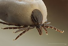
Castor bean tick, Ixodes ricinusCharacters:
  1. Ticks are ectoparasites (external parasites), living by hematophagy on the blood of mammals, birds, and sometimes reptiles and amphibians.
  2. Ticks, like mites, have bodies which are divided into two primary sections: the anterior capitulum (or gnathosoma), which contains the head and mouthparts; and the posterior idiosoma which contains the legs, digestive tract, and reproductive organs.
  3. Ticks satisfy all of their nutritional requirements on a diet of blood, a practice known as hematophagy.
  4. Like all arachnids, ticks have eight legs. The legs of Ixodidae and Argasidae are similar in structure. Each leg is composed of six segments: the coxa, trochanter, femur, patella, tibia, and tarsus. Each of these segments are connected by muscles which allow for flexion and extension, however the coxae have limited lateral movement. When not being used for walking, the legs remain tightly folded against the body.
Sientific classification
Kingdom: Animalia
Phylum: Arthropoda
Class: Arachnida
Subclass: Acari
Superorder: Parasitiformes
Order: Ixodida
Leach, 1815
Superfamily: Ixodoidea
Leach, 1815


Classify & give characters of Sarcopets (Mits)


Kingdom: Animalia

Phylum: Arthropoda

Class: Arachnida

Subclass: Acari


Family: Sarcoptidae

 Genus: Sarcoptes


Characters:
01.                     Sarcoptic Mange Mites are tiny arachnids (cousins of ticks and spiders) that are parasites of mammals. They cause the disease known as "mange" or "scabies."
02.                     These mites are tiny, only 1/64 of an inch long. They are pearly white in color and oval-shaped. They have spines on their bodies and legs. They have no eyes.
03.                     Sarcoptic Mange Mites spend their entire life on their hosts. The host is the animal that the mite lives on.
04.                     Sarcoptic Mange Mites are parasites of squirrels, rabbits, foxes, dogs, humans, and many other mammals.
05.                     Scarcoptic Mange Mites use small suckers on their legs to hold onto their hosts.
06.                     After mating, female mites burrow into the skin of the host. They use their jaws and front legs to cut the skin. They mites tunnel in the top layer of the skin only.
07.                     Inside the burrow, the female will lay eggs. She lays two or three eggs each day, for up to two months.
08.                     Mite larvae hatch from the eggs in three or four days. They immediately crawl out of the burrow onto the surface of the skin. The will stay here, using the host's hair as shelter. Both larvae and adult mites eat skin cells from their hosts.
09.                     Once a larva has eaten enough, it will molt (shed its skin). After it molts twice, it has become an adult.
10.                     Adult mange mites mate on the surface of the host's skin.
11.                     The disease caused by these mites, called "mange" or "scabies," starts with a substance from the mites' bodies. This substance causes an allergic reaction in the host's skin, and it becomes very itchy.
12.                     When the host scratches itself, it makes wounds that become infected by bacteria. The scratching also causes hair to fall out and animals with mange will often have bald patches.

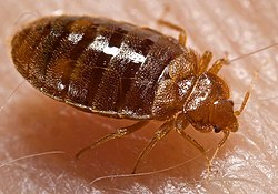
Classify & Give 5 characters of Bedbug (Cimex)

Bed bug
Cimex lectularius
Scientific classification
Kingdom: Animalia
Phylum: Arthropoda
Class: Insecta
Order: Hemiptera
Suborder: Heteroptera
Infraorder: Cimicomorpha
Superfamily: Cimicoidea
Family: Cimicidae
Latreille, 1802
Subfamilies, Genera & Species

Give a brief description of Hirudo

Medicinal leech - Hirudo medicinalis Linnaeus, 1758


Classification 

Kingdom: Animalia

Phylum: Annelida

Class: Clitellata

Order: Arhynchobdellida

Family: Hirudinidae

Genus: Hirudo

Species: H. medicinalis 

Geographical Distribution

Once abundant in Europe from Ireland to the Ural mountains (western Russia) and from southern Scandinavia to the Mediterranean. It is still present in one or more localities in 24 European countries although it is threatened in at least 12 of these. Its status is uncertain in Portugal, Sicily and Turkey and it has been extinct in Ireland for at least 100 years. It was imported into North America for medicinal purposes but no recent records have shown its presence in the wild. Present in small numbers in Britain particularly in rural areas such as New Forest, the Lake District, South Wales, Anglesey and the west of Scotland. The largest population in Britain is thought to be in Dungeness, Kent where a mark-release-recapture experiment indicated a self-sustaining population of 6,000 - 12,000 leeches in 1985.

Ecological and Habitat Requirements

Hirudo medicinalis typical habitats consist of shallow, eutrophic ponds or lakes with dense stands of macrophytes and high summer temperatures. High temperature requirements have important implications for the distribution of the medicinal leech, for example, it cannot survive in many lakes and most of the tarns in the Lake District due to the relatively low temperatures. The average temperatures for 10%, 50% and 90% activity in the medicinal leech are 11.9˚C, 19.0˚C and 22.9˚C respectively. The minimum temperature threshold for swimming activity is 5-9˚C. In a temperature gradient of 7-43˚C, studies have shown that 21˚C is the preferred temperature. The optimum temperature ranges for growth and breeding are 22-25˚C and 25.5-27.5˚C respectively with the upper lethal range being 39-43.5˚C.

Food and Feeding

H. medicinalis is the only species known to attack humans. Like other Gnathobdellid leeches, it has a short muscular pharynx usually with toothed jaws. It feeds almost exclusively on the blood of mammals but other hosts include amphibians and small fish. A single leech can take two to five times its own weight of blood in a meal, which is digested slowly over several months. For example, a leech weighing 128mg dry weight took 640mg dry weight of blood in one meal, took about 200 days to digest this meal and survived for a further 100 days without another meal. Therefore, one meal can sustain a leech for a year. The bacterium Aeromonas hydrophila is a normal symbiont in the gut of medicinal leeches which produces enzymes necessary to digest the blood ingested by the leech. Although it takes only about half an hour for a leech to become satiated on a mammalian host, the host can move a considerable distance in this time and therefore it is thought that in nature, leeches frequently detach from the host long before they are satiated to ensure they do not become isolated from their habitat.

Life Cycle Summary

Leeches are protandrous hermaphrodites and tend to cross fertilise by taking up a head-to-tail position with another leech. H. medicinalis has an eversible penis, a single male and a single female pore, a single pair of ovaries and between 10 and 100 pairs of testes. Matings occur in summer and sperm can be stored so there can be a delay of 1-9 months between copulation and cocoon deposition. Segments near the middle of the body are modified in mature worms to form a clitellum that secretes a cocoon for the eggs. Each sclerotized cocoon is about 10mm long, either oval or spherical in shape with the wall consisting of an inner smooth layer and an outer spongy layer and contains between 5 and 15 eggs. They are laid in a damp place just above the water line on the shore or bank of lakes or ponds. Cocoons are normally found in July and August and hatching time varies from 4 to 10 weeks depending on temperature. In laboratory conditions, H. medicinalis lay 1-7 cocoons in which 3-30 eggs develop and each individual can produce 2 broods per year under optimum conditions. Newly hatched leeches weigh approximately 0.12-0.18g live weight and this increases to 0.5-0.6g by end of first year, about 1.4g in second year and 2.4g in third year. It is thought that they take at least 2 years to reach sexual maturity in the field and slow-growing leeches may not breed until 3 or 4 years of age. 

Life Stages


Life Stage: Adult

Definitive Host: Mammals (including humans), amphibians and fish.
Site Within Host: Ectoparasitic.

Host Habitat: Freshwater ponds and lakes.

Free Living Environment: Freshwater ponds and lakes (also comes onto the shore to lay cocoons containing eggs).

Reproduction Capacity: Sexually mature.

Morphology: Red-yellow longitudinal stripes on the dorsal surface forming part of a fairly ornate pattern, anus small and scarcely visible, each jaw is armed with a single row of numerous sharp teeth capable of piercing human skin.

Economic Value

Raised commercially in ponds in the 18th and 19th centuries especially in France, Hungary and Russia, they were used occasionally as economic barometers but primarily for blood-letting (phlebotomy), to drain haematomas and to enhance the success of tissue transplants. Blood-letting was a common medical practice and an estimated 25 million leeches were used in France in 1846 and a further 7 million in London hospitals in 1863. This large trade in medicinal leeches meant the indigenous supply had to be supplemented by large importations. H. medicinalis is also a popular subject for laboratory studies of morphology, physiology and behaviour and is now in high demand for experimental biology (2000 are used in the US per year), plastic surgery, the provision of pharmaceutical products and neurobiology (thought to be a model organism for studies as they have exceptionally large nerve cells and supporting glial cells). The saliva of H. medicinalis contains hirudin, the most powerful anticoagulant known. Hirudin is an acidic polypeptide with a molecular weight of about 7000. The collection of hirudin led to the destruction of a large number of leeches but now as a result of cloning and expression of a recombinant gene for hirudin in yeast and bacteria, fewer animals are destroyed. Other important compounds in the saliva are histamine (causes vasodilation of blood vessels in host), hyaluronidase (breakdown of host tissues increases permeability) and bdellin & eglin (basis of anti-inflammatory response in host). 

Threats

In the past, over-collection of H. medicinalis for blood-letting has been blamed for the rarity of these leeches but in fact these leeches were frequently discarded into the nearest ditch or pond after use and this practice may have helped the survival of the species. However, more recent collections of H. medicinalis for experimental biology, medical and pharmaceutical needs pose a more serious threat as these leeches are destroyed. Today there is still a large trade in medicinal leeches which are still exported from France, Italy, Portugal, Hungary, Romania, Yugoslavia, Turkey and Greece (where they are supposed to be legally protected). The change or loss of prime medicinal leech habitat has also helped to hasten their decline. The alteration of many natural shallow ponds to deeper fishponds more suitable for trout and the general loss of wetlands, especially flushes and marshes, have not only reduced leech habitat but has also led to a decline in amphibians, an important host for the medicinal leech. Change in habitat, as well as their destruction, must be considered in conservation management for both the leech and their potential host species. The rearing of captive leeches is encouraged in order to reduce the number of individuals removed from the wild and the establishment of a leech farm in Swansea in 1984 was a welcome development. Its aim was to meet the world demand for leeches for use in research and medicine and today it provides the international market with around 15,000 leeches per year.

Legal Protections

Although H. medicinalis has been removed from the UK BAP list as it is not considered to meet the UK priority species criteria, it is included in the 1996 International Union for the Conservation of Nature (IUCN) invertebrate red data book as a low risk, near threatened species, and also in the Finnish red data book. It was also added in March 1988 to the list of species protected under the Wildlife and Countryside Act (1981) making it an offence to kill, injure, take, posses or sell (alive or dead) H. medicinalis from the wild or to damage, destroy or obstruct access to their natural habitat.